What Is Stretching?
Stretching is something that we all do. You’ll see dogs do it when they get out of bed. They’ll stretch, then yawn. You’ll see babies do it. It’s a natural process of being a human and animal.
Many people make the incorrect assumption that when muscles are tight, you need to stretch them, and by doing so, you will gain flexibility and muscles physically become stronger which is incorrect. Our muscles don’t really stretch. What we’re doing is we’re stretching our connective tissue and neurologically retraining our muscles.
If you feel that you are not flexible, it is not because your muscles are tight. It is because your brain is not allowing your muscles to move along to their full length. Your nervous system is inhibiting your muscles from changing their shape as a safety precaution. Flexibility is about gaining control over your muscles; it’s not about stretching them.
One of the aspects that proper stretching is doing, is giving your nervous system a signal that you are fully in control, that you’re strong enough to protect yourself in that lengthened position and that this position is not harming you. One of the problems, when you’re in this lengthened position, is that your muscles are in a very weak and vulnerable place. They are mechanically disadvantaged to contract. Building control and moving without pain in these weakened ranges will actually increase your flexibility. When you stretch over the course of 6 to 12 weeks, what you are in fact doing is just increasing the tolerance to a certain position. You are perceiving less threat in that new range.
Let’s learn more about the physiologic aspects of stretching. Then we’ll look at how you can pick the right type of stretching to treat your conditions, problems or training.
Stretching
Stretching of any kind helps improve recovery and performance. Choosing the right type of stretch depends on two things:
- The type of activity you are doing
- Whatever gives you the best results
A good rule of thumb is you should enjoy stretching. You should not hurt while doing it and you should feel better, not worse afterwards.
Physiology of Stretching
There are two major components. There’s the muscle component and there’s a connective tissue component. Let’s talk about the muscle.
Your muscles are made up of myofibrils which are made up of actin myosin. You have these interlinking fibers and when your muscles contract they go together. When your muscles relax they come apart, and your nervous system controls this contraction and relaxation. Your nervous system also controls what level these fibers are interlinking. At 130% of a stretch from a relaxed position these fibers, you start to experience weakness. At 150% of a stretch, you start to actually tear the muscles. Your body doesn’t want your muscles to tear. When they are over-extended, it has this neurological system called your proprioceptive system. The proprioceptive system perceives where your muscle is in space and how much length each muscle has.
The Musculoskeletal System
Together, muscles and bones comprise what is called the musculoskeletal system of the body. The bones provide posture and structural support for the body and the muscles provide the body with the ability to move (by contracting and thus generating tension). The point where bones connect to one another is called a joint, and this connection is made mostly by ligaments (along with the help of muscles). Muscles are attached to the bone by tendons. Bones, tendons, and ligaments do not possess the ability (as muscles do) to make your body move. Muscles are very unique in this respect.
Muscle Composition
At the highest level, the (whole) muscle is composed of many strands of tissue called fascicles. These are the strands of muscle that we see when we cut red meat or poultry. Each fascicle is composed of fasciculi which are bundles of muscle fibers. The muscle fibers are in turn composed of tens of thousands of thread-like myofybrils, which can contract, relax, and elongate (lengthen). The myofybrils are (in turn) composed of up to millions of bands laid end-to-end called sarcomeres. Each sarcomere is made of overlapping thick and thin filaments called myofilaments. The thick and thin myofilaments are made up of contractile proteins, primarily actin and myosin.
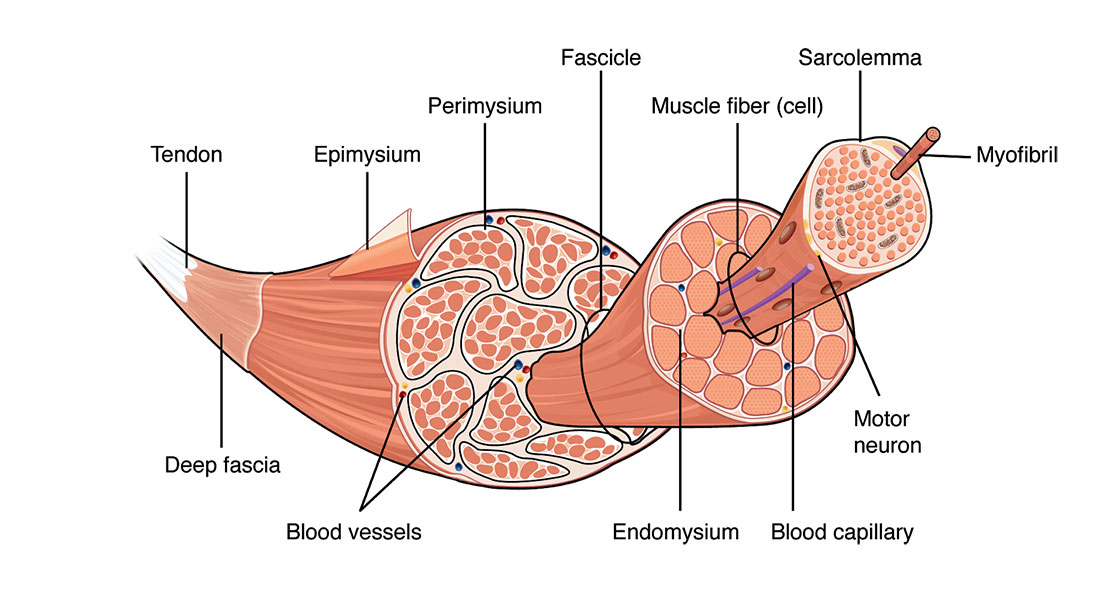
Image provided by Oregon State University, Open Educational Resources Unit
How Muscles Contract
Nerves connect the spinal column to the muscle. The place where the nerve and muscle meet is called the neuromuscular junction. When an electrical signal crosses the neuromuscular junction, it is transmitted deep inside the muscle fibers. Inside the muscle fibers, the signal stimulates the flow of calcium which causes the thick and thin myofilaments to slide across one another. When this occurs, it causes the sarcomere to shorten, which generates force. When billions of sarcomeres in the muscle shorten all at once it results in a contraction of the entire muscle fiber.
Interestingly muscles, when they contract, are always 100%. Muscle fibers are unable to vary the intensity of their contraction relative to the load against which they are acting.What happens is when we’re increasing the intensity of muscle contractions, it is actually the nervous system that is recruiting more and different types of muscle fibers to contract.
This is important when we look into stretching, because one of the things that happens is when we stretch the muscles, we’re not actually stretching all the muscle fibers. We are stretching some of the muscle fibers and a way to increase the stretch is actually by contracting the muscles which engages muscle fibers.
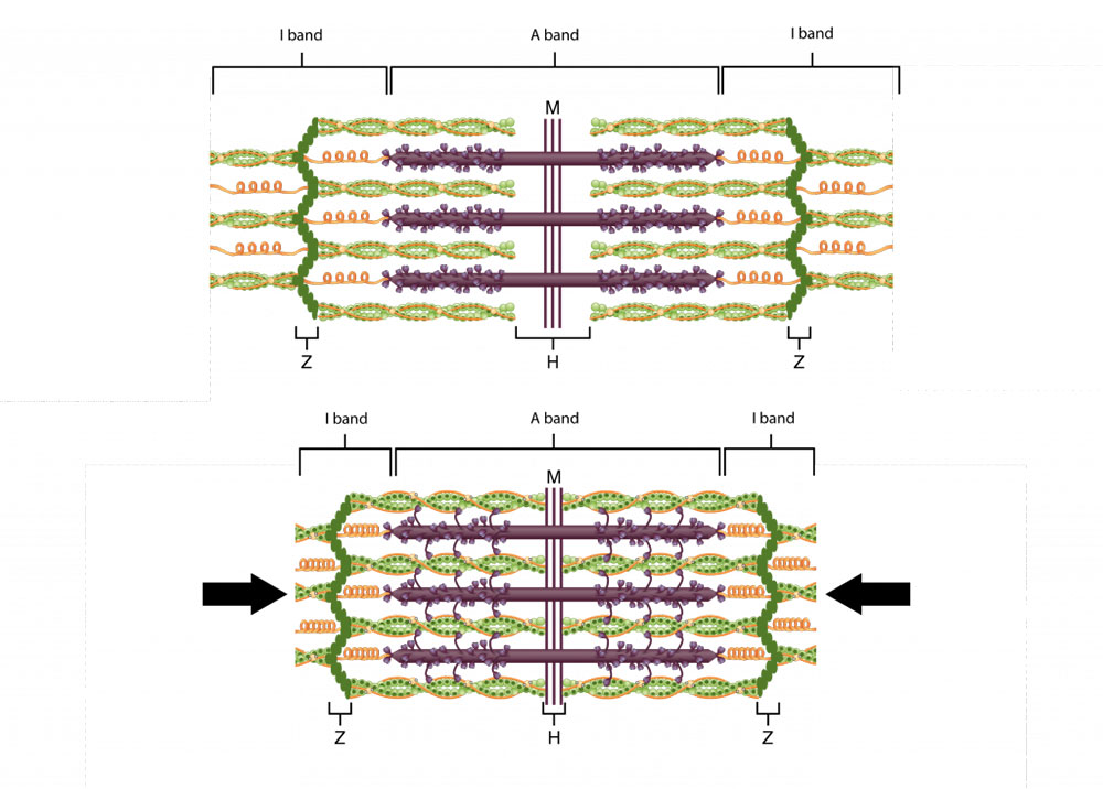
Image provided by Oregon State University, Open Educational Resources Unit
Fast and Slow Muscle Fibers
The energy which produces the calcium flow in the muscle fibers comes from mitochondria, the part of the muscle cell that converts glucose (blood sugar) into energy. Different types of muscle fibers have different amounts of mitochondria. The more mitochondria in a muscle fiber, the more energy it is able to produce.
Muscle fibers are categorized into slow-twitch fibers and fast-twitch fibers. Slow-twitch fibers (also called Type 1 muscle fibers) are slow to contract, but they are also very slow to fatigue. Fast-twitch fibers are very quick to contract and come in two varieties: Type 2A muscle fibers which fatigue at an intermediate rate, and Type 2B muscle fibers which fatigue very quickly. Slow-twitch fibers are also smaller in diameter than fast-twitch fibers and have increased capillary blood flow around them. Because they have a smaller diameter and an increased blood flow, the slow-twitch fibers are able to deliver more oxygen and remove more waste products from the muscle fibers (which decreases their “fatigability”).
Dark meat is dark because it has a greater number of slow-twitch muscle fibers and hence a greater number of mitochondria, which are dark. White meat consists mostly of muscle fibers which are at rest much of the time but are frequently called on to engage in brief bouts of intense activity. This muscle tissue can contract quickly but is fast to fatigue and slow to recover. White meat is lighter in color than dark meat because it contains fewer mitochondria.
These three muscle fiber types (Types 1, 2A, and 2B) are contained in all muscles in varying amounts. Muscles that need to be contracted much of the time (like the heart) have a greater number of Type 1 (slow) fibers. When a muscle first starts to contract, it is primarily Type 1 fibers that are initially activated, then Type 2A and Type 2B fibers are activated (if needed) in that order. The fact that muscle fibers are recruited in this sequence is what provides the ability to execute brain commands with such fine-tuned muscle responses. It also makes the Type 2B fibers difficult to train because they are not activated until most of the Type 1 and Type 2A fibers have been recruited.
A side note with endurance athletes, what you’ll find is you don’t want all your muscles to contract all the time. This would fatigue the muscles too quickly and it actually trains the nervous system to fire those muscle fibers in a rotational pattern to increase endurance. So while some muscles are contracting, some are resting.
On the other hand, power lifters and explosive athletes, you will actually want as much muscle contraction as you can, so you’re neurologically training yourself to fire all your muscles at once.
Connective Tissue
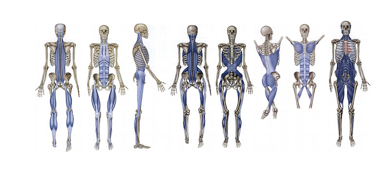
The connective tissue is your tendons, ligaments and fascia. Ligaments are what connect bone to bone and your tendons are what connects your muscles to your bones. We don’t want to stretch ligaments. They are meant to hold your bones together. Outside of ligaments and tendons we have fascia, a connective tissue that surrounds every fiber of your muscle. Not only around the muscle, but also inside the muscle. Every single little muscle fiber has a little mini envelope of connective tissue around it. The whole muscle is wrapped in this multidimensional tube of connective tissue. The connective tissue doesn’t stop there, it goes between each muscle. This connection transmits the force from one muscle to the next. We used to think the force that a muscle exerts, presses on the tendon and then presses on the bone. That’s actually not the case; a large component of the force that a muscle exerts, goes laterally to the connective tissue and around it, and then to the muscles beside it. Connective tissue distributes the force throughout the limb. Connective tissue doesn’t just stop at the musculus, it surrounds every single organ in the body. This is what we call a matrix of connective tissue. It is the scaffold that gives our body shape. Connective tissue is the common denominator throughout the entire body. Fascia connective tissue literally connects us. It is what holds you together and provides your shape and posture. It is a state of structural and functional continuity between all of the body’s hard and soft tissues, Without fascia, our muscles would be like a jelly substance without much form at all.
This structure it provides is not passive. Fascial tissue contains contractile cells which influence musculoskeletal dynamics. Fascia is one of our most important perceptual tissues The fascia contains sensitive nerves that convey proprioception (joint position sense) fascial tissue is one of our richest sensory organs. A myriad of tiny unmyelinated ‘free’ nerve endings are found almost everywhere in fascial tissues, but particularly in bone and visceral connective tissues.
Connective tissue is composed of a base substance and two kinds of protein based fiber. The two types of fiber are:
- Collagenous connective tissue (consists mostly of collagen and provides tensile strength)
- Elastic connective tissue (consists mostly of elastin and provides elasticity)
The base substance is called mucopolysaccharide and acts as both a lubricant (allowing the fibers to easily slide over one another), and as a glue (holding the fibers of the tissue together into bundles). The more elastic connective tissue there is around a joint, the greater the range of motion in that joint.
Fascia, when healthy, forms a gliding interface with underlying muscle allowing free excursion of the muscle under the relatively immobile skin. When we’re stretching the connective tissue, we’re actually stretching the more elastic quality fibers.
Connective tissue is also responsive to heat. That’s why when you do a warm up, you increase your body’s temperature, or when you take a hot shower, you feel more flexible as this tissue is more fluid. Just like every other tissue in your body, your fascia is made of water. It works better, moves better and feels better when it’s wet. So, drink more water! We’re not just stretching muscle but stretching this connective tissue that flows throughout the body that goes into our organs.
Why It’s Important To Stretch Our Connective Tissue
When you stretch connective tissue it stimulates anti-inflammatory agents in the body. So stretching can decrease inflammation, specifically when you’re stretching connective tissue. One of the benefits when we think about yoga and tai chi as being helpful with arthritis and other inflammation, is that we are stretching this connective tissue.
Acute inflammation is accompanied from its outset by the release of specialized pro-resolving mediators (SPMs), including resolvins (Resolvins are specialized pro-resolving mediators derived from omega-3 fatty acids), that orchestrate the resolution of local inflammation.
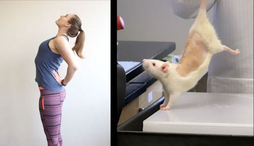
In rats with subcutaneous inflammation of the back, carrageenan was induced. Stretching for 10 minutes twice daily reduced inflammation and improved pain, two weeks after carrageenan injection.
Furthermore, subcutaneous resolving injection mimicked the effect of stretching. In ex vivo experiments, stretching of connective tissue reduced the migration of neutrophils and increased tissue RvD1 concentration. These results demonstrate a direct mechanical impact of stretching on inflammation-regulation mechanisms within connective tissue.
In a recent ultrasound study, human subjects with chronic low back pain had altered connective tissue structure compared to human subjects without low back pain, suggesting the presence of inflammation and/or fibrosis in the low back pain subjects. Mechanical input in the form of static tissue stretch has been shown in vitro and in vivo to have anti-inflammatory and anti-fibrotic effects.
Showed that gentle daily stretching for 10 minutes can reduce local connective tissue inflammation and fibrosis. Because mechanical factors within the stroma can influence the tumor microenvironment, they suggest a link between immune exhaustion, inflammation resolution and tumor growth. Stretching is a gentle, non-pharmacological intervention that could become an important component of cancer treatment and prevention.
What Happens When You Stretch
The stretching of a muscle fiber begins with the sarcomere, the basic unit of contraction in the muscle fiber. As the sarcomere contracts, the area of overlap between the thick and thin myofilaments increases. As it stretches, this area of overlap decreases, allowing the muscle fiber to elongate. Once the muscle fiber is at its maximum resting length (all the sarcomeres are fully stretched), additional stretching places force on the surrounding connective tissue. As the tension increases, the collagen fibers in the connective tissue align themselves along the same line of force as the tension. Hence when you stretch, the muscle fiber is pulled out to its full length sarcomere by sarcomere, and then the connective tissue takes up the remaining slack. When this occurs, it helps to realign any disorganized fibers in the direction of the tension. This realignment is what helps to rehabilitate scarred tissue back to health.
When a muscle is stretched, some of its fibers lengthen, but other fibers may remain at rest. The current length of the entire muscle depends upon the number of stretched fibers (similar to the way that the total strength of a contracting muscle depends on the number of recruited fibers contracting). The more fibers that are stretched, the greater the length developed by the stretched muscle.
Proprioceptors
The nerve endings that relay all the information about the musculoskeletal system to the central nervous system are called proprioceptors. Proprioceptors (also called mechanoreceptors) are the source of all proprioception: the perception of one’s own body position and movement. The proprioceptors detect any changes in physical displacement (movement or position) and any changes in tension, or force, within the body. They are found in all nerve endings of the joints, muscles, and tendons. The proprioceptors related to stretching are located in the tendons and in the muscle fibers.
There’s two components of that proprioceptive system that we will focus on:
- Muscle spindle fibers (lMonitor Muscle Length)
- Golgi tendon (Monitor Muscle Tension)
These are basically the nerves that talk to the muscles (muscle spindle fibers) and the nerves that talk to the tendons (golgi tendon).
The stretch receptors which are sensitive to the change in muscle length and the rate of change in muscle length. When muscles contract, it places tension on the tendons where the golgi tendon organ is located. The golgi tendon organ is sensitive to the change in tension and the rate of change of the tension. Tension that is caused by muscular contraction, but not passive stretch.
What happens is when there’s a quick pulling apart the muscle spindle fibers in the muscles, will send a neurological signal and actually cause the muscle to contract.
When there’s injury, obviously the body feels like it can’t stretch very far and so it keeps those muscle spindles close together. So when you’re stretching you don’t want to have pain because that is reinforcing a contraction signal.
You want a stretch that’s safe. What happens is as you hold that stretch for a long period of time, usually 30 – 60 seconds. Generally you’re holding it longer because what you’re doing is you’re decreasing that signal. That stretch signal to the nervous system you’re getting the nervous system used to having those muscle fibers being held at that length, what the body perceives a weakened state or a dangerous state. So you put the muscle fibers in the stretch and then you hold it there so the nervous system can adapt.
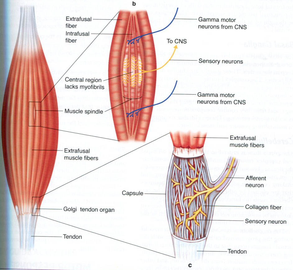
Image provided by neurones.co.uk
The Stretch Reflex (Myotatic Reflex)
When the muscle is stretched, so is the muscle spindle. The muscle spindle records the change in length (and how fast) and sends signals to the spine which convey this information. This triggers the stretch reflex (also called the myotatic reflex) which attempts to resist the change in muscle length by causing the stretched muscle to contract. The more sudden the change in muscle length, the stronger the muscle contractions will be (plyometric, or “jump”, training is based on this fact). This basic function of the muscle spindle helps to maintain muscle tone and to protect the body from injury.
This is the reflex the doctors are checking when they hit your knee with the hammer. The hammer stretches the muscle quickly and causes a contraction.
The stretch reflex has both a dynamic component and a static component. The static component of the stretch reflex persists as long as the muscle is being stretched. The dynamic component of the stretch reflex (which can be very powerful) lasts for only a moment and is in response to the initial sudden increase in muscle length. The reason that the stretch reflex has two components is because there are actually two kinds of intrafusal muscle fibers: nuclear chain fibers, which are responsible for the static component; and nuclear bag fibers, which are responsible for the dynamic component.
Nuclear chain fibers are long and thin, and lengthen steadily when stretched. When these fibers are stretched, the stretch reflex nerves increase their firing rates (signaling) as their length steadily increases. This is the static component of the stretch reflex.
Nuclear bag fibers bulge out at the middle, where they are the most elastic. The stretch-sensing nerve ending for these fibers is wrapped around this middle area, which lengthens rapidly when the fiber is stretched. The outer-middle areas, in contrast, act like they are filled with viscous fluid; they resist fast stretching, then gradually extend under prolonged tension. So, when a fast stretch is demanded of these fibers, the middle takes most of the stretch at first; then, as the outer-middle parts extend, the middle can shorten somewhat. So the nerve that senses stretching in these fibers fires rapidly with the onset of a fast stretch, then slows as the middle section of the fiber is allowed to shorten again. This is the dynamic component of the stretch reflex: a strong signal to contract at the onset of a rapid increase in muscle length, followed by slightly “higher than normal” signaling which gradually decreases as the rate of change of the muscle length decreases.
One of the reasons for holding a stretch for a prolonged period of time is that as you hold the muscle in a stretched position, the muscle spindle habituates (becomes accustomed to the new length) and reduces its signaling. Gradually, you can train your stretch receptors to allow greater lengthening of the muscles.
Some sources suggest that with extensive training, the stretch reflex of certain muscles can be controlled so that there is little or no reflex contraction in response to a sudden stretch. While this type of control provides the opportunity for the greatest gains in flexibility, it also provides the greatest risk of injury if used improperly. Only consummate professional athletes and dancers at the top of their sport (or art) are believed to actually possess this level of muscular control.
The Lengthening Reaction (Inverse Myotatic Reflex)
When muscles contract, they produce tension at the point where the muscle is connected to the tendon, where the golgi tendon organ is located. The golgi tendon organ records the change in tension, and the rate of change of the tension, and sends signals to the spine to convey this information. When this tension exceeds a certain threshold, it triggers the lengthening reaction which inhibits the muscles from contracting and causes them to relax. Other names for this reflex are the inverse myotatic reflex, autogenic inhibition, and the clasped-knife reflex. This basic function of the golgi tendon organ helps to protect the muscles, tendons, and ligaments from injury. The lengthening reaction is possible only because the signaling of the golgi tendon organ to the spinal cord is powerful enough to overcome the signaling of the muscle spindles telling the muscle to contract.
So if you’re lifting something heavy and the tendon feels like it’s going to get damaged, it’ll turn the muscles off. You’ll see that with hamstring strains and other similar injuries.
It was first thought GTOs only had a protective function ,but it is now known that golgi tendon signal muscle tension continuously providing precise information about muscle force, that the reflex pathway has multisensory inputs that may allow precise control of muscle forces for fine activities.
We have the golgi tendons and we have the muscle spindle fibers that inversely talk to the nervous system, where one is more inhibiting and the other is causing contractions but they’re working together as a team. The nervous system is controlling the muscles telling it to contract. The nervous system is listening to the muscles, how far are they expanding and how much tension is on the tendons. It is infoming how much contraction is in the muscles. When we stretch we want to let the nervous system know, that we’re safe and that we’re okay. That there’s no pain and in this stretch position it’s fine. The nervous system will basically reset those muscle fibers. It’s not like your muscles are tight, it’s not like your muscles are loose. It’s more about what your body perceives is safe.
With injury, the body doesn’t perceive that it is safe and it will cause a muscle contraction. Basically it’s causing a cast so that you have less range of motion. It’s a spasm of the muscle and so one of the things that we do is let the body know that this motion is safe. Two key points to take away:
- You want to hold a muscle for a long time to help reinforce a signal that this is a safe position. Let the nervous system adjust to that.
- You want to avoid pain when you do a stretch, because that’s reinforcing that this is not safe and then also we have the contraction phase to increase more fibers.
Another reason for holding a stretch for a prolonged period of time is to allow this lengthening reaction to occur, thus helping the stretched muscles to relax. It is easier to stretch, or lengthen, a muscle when it is not trying to contract.
Clinical Note
Upper motoneuron lesions which damage the descending pathways down to the spinal cord may cause increase in muscle tone, partly because motoneurons respond more to muscle spindle afferent inputs. This causes increased resistance to passive movement (that the patient doesn’t initiate), called spasticity, which is associated with another neurological sign, the clasp-knife response, in which the spastic muscle initially resists passive movement strongly, and then suddenly yields.
Reciprocal Inhibition
Another thing that the nervous system does is called reciprocal inhibition. When one muscle contracts the other muscle has to relax. Our muscles work together in pairs.
Agonists
These muscles cause the movement to occur. They create the normal range of movement in a joint by contracting. Agonists are also referred to as prime movers since they are the muscles that are primarily responsible for generating the movement.
Antagonists
These muscles act in opposition to the movement generated by the agonists and are responsible for returning a limb to its initial position.
When an agonist contracts, in order to cause the desired motion, it usually forces the antagonists to relax. This phenomenon is called reciprocal inhibition because the antagonists are inhibited from contracting. This is sometimes called reciprocal innervation but that term is really a misnomer since it is the agonists which inhibit (relax) the antagonists. The antagonists do not actually innervate (cause the contraction of) the agonists.
Such inhibition of the antagonistic muscles is not necessarily required. In fact, co-contraction can occur. When you perform a sit-up, one would normally assume that the stomach muscles inhibit the contraction of the muscles in the lumbar, or lower, region of the back. In this particular instance however, the back muscles (spinal erectors) also contract. This is one reason why sit-ups are good for strengthening the back as well as the stomach.
As an example, when you flex your knee, your hamstring contracts, and, to some extent, so does your gastrocnemius (calf) and lower buttocks. Meanwhile, your quadriceps are inhibited (relaxed and lengthened somewhat) so as not to resist the flexion. In this example, the hamstring serves as the agonist, or prime mover; the quadricep serves as the antagonist; and the calf and lower buttocks serve as the synergists. Agonists and antagonists are usually located on opposite sides of the affected joint (like your hamstrings and quadriceps, or your triceps and biceps), while synergists are usually located on the same side of the joint near the agonists. Larger muscles often call upon their smaller neighbors to function as synergists.
The following is a list of commonly used agonist/antagonist muscle pairs:
- pectorals/latissimus dorsi (pecs and lats)
- anterior deltoids/posterior deltoids (front and back shoulder)
- trapezius/deltoids (traps and delts)
- abdominals/spinal erectors (abs and lower-back)
- left and right external obliques (sides)
- quadriceps/hamstrings (quads and hams)
- shins/calves
- biceps/triceps
- forearm flexors/extensors
One of the things that you can do with your stretching is engage that process. For example, if you’re stretching your calf and you actively lift your toe, you are contracting your anterior tibialis which will reciprocally inhibit your calf muscles. It will help the nervous system let those muscles go. It is easier to stretch a muscle that is relaxed than to stretch a muscle that is contracting. By taking advantage of the situations when reciprocal inhibition does occur, you can get a more effective stretch by inducing the antagonists to relax during the stretch due to the contraction of the agonists.
When we stretch muscles, what we’re working on is the nervous system. We’re not really stretching, we’re just retraining. We don’t actually stretch the muscles, we’re relaxing the muscles.
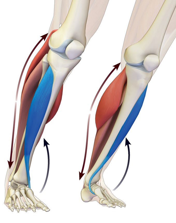
Image provided by bandhayoga.com
Factors Limiting Flexibility
According to Gummerson, flexibility (he uses the term mobility) is affected by the following factors:
Internal influences
- the type of joint (some joints simply aren’t meant to be flexible)
- the internal resistance within a joint
- bony structures which limit movement
- the elasticity of muscle tissue (muscle tissue that is scarred due to a previous injury is not very elastic)
- the elasticity of tendons and ligaments (ligaments do not stretch much and tendons should not stretch at all)
- the elasticity of skin (skin actually has some degree of elasticity, but not much)
- the ability of a muscle to relax and contract to achieve the greatest range of movement
- the temperature of the joint and associated tissues (joints and muscles offer better flexibility at body temperatures that are 1 to 2 degrees higher than normal)
External influences
- the temperature of the place where one is training (a warmer temperature is more conducive to increased flexibility)
- the time of day (most people are more flexible in the afternoon than in the morning, peaking about 2:30pm-4pm)
- the stage in the recovery process of a joint (or muscle) after injury (injured joints and muscles will usually offer a lesser degree of flexibility than healthy ones)
- age (pre-adolescents are generally more flexible than adults)
- gender (females are generally more flexible than males)
- one’s ability to perform a particular exercise (practice makes perfect)
- one’s commitment to achieving flexibility
- the restrictions of any clothing or equipment
Some sources also suggest that water is an important dietary element with regard to flexibility. Increased water intake is believed to contribute to increased mobility, as well as increased total body relaxation.
How Connective Tissue Affects Flexibility
The resistance to lengthening that is offered by a muscle is dependent upon its connective tissues: When the muscle elongates, the surrounding connective tissues become more taut. Also, inactivity of certain muscles or joints can cause chemical changes in connective tissue which restrict flexibility. According to M. Alter, each type of tissue plays a certain role in joint stiffness: “The joint capsule (i.e., the saclike structure that encloses the ends of bones) and ligaments are the most important factors, accounting for 47 percent of the stiffness, followed by the muscle’s fascia (41 percent), the tendons (10 percent), and skin (2 percent)”.
M. Alter goes on to say that efforts to increase flexibility should be directed at the muscle’s fascia however. This is because it has the most elastic tissue, and because ligaments and tendons (since they have less elastic tissue) are not intended to be stretched very much at all. Overstretching them may weaken the joint’s integrity and cause destabilization (which increases the risk of injury).
When connective tissue is overused, the tissue becomes fatigued and may tear, which also limits flexibility. When connective tissue is unused or under used, it provides significant resistance and limits flexibility. The elastin begins to fray and loses some of its elasticity, and the collagen increases in stiffness and in density. Aging has some of the same effects on connective tissue that lack of use has.
How Aging Affects Flexibility
With appropriate training, flexibility can, and should, be developed at all ages. This does not imply, however, that flexibility can be developed at the same rate by everyone. In general, the older you are, the longer it will take to develop the desired level of flexibility. Hopefully, you’ll be more patient if you’re older.
According to M. Alter, the main reason we become less flexible as we get older is a result of certain changes that take place in our connective tissues. As we age, our bodies gradually dehydrate to some extent. It is believed that “stretching stimulates the production or retention of lubricants between the connective tissue fibers, thus preventing the formation of adhesions”. Hence, exercise can delay some of the loss of flexibility that occurs due to the aging process.
M. Alter further states that some of the physical changes attributed to aging are the following:
- An increased amount of calcium deposits, adhesions, and cross-links in the body.
- An increase in the level of fragmentation and dehydration.
- Changes in the chemical structure of the tissues.
- Loss of suppleness due to the replacement of muscle fibers with fatty, collagenous fibers.
This does not mean that you should give up trying to achieve flexibility if you are old or inflexible. It just means that you need to work harder, and more carefully, for a longer period of time when attempting to increase flexibility. Increases in the ability of muscle tissues and connective tissues to elongate (stretch) can be achieved at any age.
Strength and Flexibility
Does stretching increase sports performance? The answer is pretty much no, but more specifically it may be depending on the activity. Try doing a static stretch, which is basically just stretching without motion. What you’re doing here is inhibiting muscles and causing the muscles to relax. If you then go from that relaxed muscle state to a dynamic activity, like a vertical jump or a deadlift you actually will see a decrease in muscle recruitment. This makes sense because you’re doing a stretch you’re telling the nervous system to relax to turn off the muscles and then you go immediately into trying to tell the nervous system to contract all the muscles at once. You’ll see a decrease in performance. This neurological decrease in performance is very temporary, So if you did a static stretch and then you ran upstairs for 10 seconds, and then you did a vertical jump, you would mitigate all that decrease in performance, which is important to realize.
The other component to that is you are increasing performance only if you are doing a motion where you’re trying to contract the muscle in a very extremely stretched position. You find that with gymnastics, martial arts and figure skating, where you are doing most of your sports and activity in this large exaggerated range of motion. But if you’re running or jumping you’re not moving in this extreme range of motion and therefore static stretching to improve performance is not effective.
Strength training and flexibility training should go hand in hand. It is a common misconception that there must always be a trade-off between flexibility and strength. Obviously, if you neglect flexibility training altogether in order to train for strength then you are certainly sacrificing flexibility (and vice versa). However, performing exercises for both strength and flexibility need not sacrifice either one. As a matter of fact, flexibility training and strength training can actually enhance one another.
Why Bodybuilders Should Stretch
One of the best times to stretch is right after a strength workout such as weightlifting. Static stretching of fatigued muscles performed immediately following the exercise(s) that caused the fatigue, helps not only to increase flexibility, but also enhances the promotion of muscular development (muscle growth), and will actually help decrease the level of post-exercise soreness.
After you have used weights (or other means) to overload and fatigue your muscles, your muscles retain a “pump” and are shortened somewhat. This “shortening” is due mostly to the repetition of intense muscle activity that often only takes the muscle through part of its full range of motion. This “pump” makes the muscle appear bigger. The “pumped” muscle is also full of lactic acid and other by-products from exhaustive exercise. If the muscle is not stretched afterward, it will retain this decreased range of motion (it sort of “forgets” how to make itself as long as it could) and the buildup of lactic acid will cause post-exercise soreness. Static stretching of the “pumped” muscle helps it to become “looser”, and to “remember” its full range of movement.
Why Contortionists Should Strengthen
The reason for this is that flexibility training on a regular basis causes connective tissues to stretch which in turn causes them to loosen (become less taut) and elongate. When the connective tissue of a muscle is weak, it is more likely to become damaged due to overstretching, or sudden, powerful muscular contractions. The likelihood of such injury can be prevented by strengthening the muscles bound by the connective tissue.
If you also lift weights, dynamic strength training for a muscle should occur before subjecting that muscle to an intense weightlifting workout. This helps to pre-exhaust the muscle first, making it easier (and faster) to achieve the desired overload in an intense strength workout. Attempting to perform dynamic strength training after an intense weightlifting workout would be largely ineffective.
If you are working on increasing (or maintaining) flexibility then it is very important that your strength exercises force your muscles to take the joints through their full range of motion. Repeating movements that do not employ a full range of motion in the joints (like cycling, certain weightlifting techniques, and pushups) can cause shortening of the muscles surrounding the joints. This is because the nervous control of length and tension in the muscles are set at what is repeated most strongly and/or most frequently.
Over Flexibility
Once a muscle has reached its absolute maximum length, attempting to stretch the muscle further only serves to stretch the ligaments and put undue stress upon the tendons (two things that you do not want to stretch). Ligaments will tear when stretched more than 6% of their normal length. Tendons are not even supposed to be able to lengthen. Even when stretched ligaments and tendons do not tear, loose joints and/or a decrease in the joint’s stability can occur (thus vastly increasing your risk of injury).
Once you have achieved the desired level of flexibility for a muscle or set of muscles and have maintained that level for a solid week, you should discontinue any isometric or PNF stretching of that muscle until some of its flexibility is lost.
Types of Stretching
Just as there are different types of flexibility, there are also different types of stretching.
Stretches are either:
- Dynamic stretches (meaning they involve motion) affect dynamic flexibility
- Static stretches (meaning they involve no motion) affect static flexibility
Dynamic Stretching
Dynamic stretching, according to Kurz, “involves moving parts of your body and gradually increasing reach, speed of movement, or both.” Do not confuse dynamic stretching with ballistic stretching! Dynamic stretching consists of controlled leg and arm swings that take you (gently!) to the limits of your range of motion. Ballistic stretches involve trying to force a part of the body beyond its range of motion. In dynamic stretches, there are no bounces or “jerky” movements. An example of dynamic stretching would be slow, controlled leg swings, arm swings, or torso twists.
Dynamic stretching improves dynamic flexibility and is quite useful as part of your warm-up for an active or aerobic workout (such as a dance or martial-arts class).
According to Kurz, dynamic stretching exercises should be performed in sets of 8-12 repetitions. Be sure to stop when and if you feel tired. Tired muscles have less elasticity which decreases the range of motion used in your movements. Continuing to exercise when you are tired serves only to reset the nervous control of your muscle length at the reduced range of motion used in the exercise (and will cause a loss of flexibility). Once you attain a maximal range of motion for a joint in any direction you should stop doing that movement during that workout. Tired and overworked muscles won’t attain a full range of motion and the muscle’s kinesthetic memory will remember the repeated shorted range of motion, which you will then have to overcome before you can make further progress.
Active Stretching
Active stretching is also referred to as static-active stretching. An active stretch is one where you assume a position and then hold it there with no assistance other than using the strength of your agonist muscles. For example, bringing your leg up high and then holding it there without anything (other than your leg muscles themselves) to keep the leg in that extended position. The tension of the agonists in an active stretch helps to relax the muscles being stretched (the antagonists) by reciprocal inhibition.
Active stretching increases active flexibility and strengthens the agonistic muscles. Active stretches are usually quite difficult to hold and maintain for more than 10 seconds and rarely need to be held any longer than 15 seconds.
Many of the movements (or stretches) found in various forms of yoga are active stretches.
Passive Stretching
Passive stretching is also referred to as relaxed stretching, and as static-passive stretching. A passive stretch is one where you assume a position and hold it with some other part of your body, or with the assistance of a partner or some other apparatus. For example, bringing your leg up high and then holding it there with your hand. The splits is an example of a passive stretch (in this case the floor is the “apparatus” that you use to maintain your extended position).
Slow, relaxed stretching is useful in relieving spasms in muscles that are healing after an injury. Obviously, you should check with your doctor first to see if it is okay to attempt to stretch the injured muscles.
Relaxed stretching is also very good for “cooling down” after a workout and helps reduce post-workout muscle fatigue, and soreness.
Isometric Stretching
Isometric stretching is a type of static stretching (meaning it does not use motion) which involves the resistance of muscle groups through isometric contractions (tensing) of the stretched muscles. The use of isometric stretching is one of the fastest ways to develop increased static-passive flexibility and is much more effective than either passive stretching or active stretching alone. Isometric stretches also help to develop strength in the “tensed” muscles (which helps to develop static-active flexibility), and seems to decrease the amount of pain usually associated with stretching.
The most common ways to provide the needed resistance for an isometric stretch are to apply resistance manually to one’s own limbs, to have a partner apply the resistance, or to use an apparatus such as a wall (or the floor) to provide resistance.
An example of manual resistance would be holding onto the ball of your foot to keep it from flexing while you are using the muscles of your calf to try and straighten your instep so that the toes are pointed.
An example of using a partner to provide resistance would be having a partner hold your leg up high (and keep it there) while you attempt to force your leg back down to the ground.
An example of using the wall to provide resistance would be the well known “push-the-wall” calf-stretch where you are actively attempting to move the wall (even though you know you can’t).
Isometric stretching is not recommended for children and adolescents whose bones are still growing. These people are usually already flexible enough that the strong stretches produced by the isometric contraction have a much higher risk of damaging tendons and connective tissue. Kurz strongly recommends preceding any isometric stretch of a muscle with dynamic strength training for the muscle to be stretched. A full session of isometric stretching makes a lot of demands on the muscles being stretched and should not be performed more than once per day for a given group of muscles (ideally, no more than once every 36 hours).
The proper way to perform an isometric stretch is as follows:
- Assume the position of a passive stretch for the desired muscle.
- Next, tense the stretched muscle for 7-15 seconds (resisting against some force that will not move, like the floor or a partner).
- Finally, relax the muscle for at least 20 seconds.
Some people seem to recommend holding the isometric contraction for longer than 15 seconds, but according to SynerStretch (the videotape), research has shown that this is not necessary. So you might as well make your stretching routine less time consuming.
How Isometric Stretching Works
Recall from our previous discussion that there is no such thing as a partially contracted muscle fiber: when a muscle is contracted, some of the fibers contract and some remain at rest (more fibers are recruited as the load on the muscle increases). Similarly, when a muscle is stretched, some of the fibers are elongated and some remain at rest. During an isometric contraction, some of the resting fibers are being pulled upon from both ends by the muscles that are contracting. The result is that some of those resting fibers stretch.
Normally, the handful of fibers that stretch during an isometric contraction are not very significant. The true effectiveness of the isometric contraction occurs when a muscle that is already in a stretched position is subjected to an isometric contraction. In this case, some of the muscle fibers are already stretched before the contraction, and, if held long enough, the initial passive stretch overcomes the stretch reflex and triggers the lengthening reaction, inhibiting the stretched fibers from contracting. When you isometrically contracted, some resting fibers would contract and some resting fibers would stretch. Furthermore, many of the fibers already stretching may be prevented from contracting by the inverse myotatic reflex (the lengthening reaction) and would stretch even more. When the isometric contraction is completed, the contracting fibers return to their resting length but the stretched fibers would remember their stretched length and (for a period of time) retain the ability to elongate past their previous limit. This enables the entire muscle to stretch beyonds its initial maximum and results in increased flexibility.
The reason that the stretched fibers develop and retain the ability to stretch beyond their normal limit during an isometric stretch has to do with the muscle spindles: The signal which tells the muscle to contract voluntarily, also tells the muscle spindle’s (intrafusal) muscle fibers to shorten, increasing sensitivity of the stretch reflex. This mechanism normally maintains the sensitivity of the muscle spindle as the muscle shortens during contraction. This allows the muscle spindles to habituate (become accustomed) to an even further-lengthened position.
PNF Stretching
PNF stretching is currently the fastest and most effective way known to increase static-passive flexibility. PNF is an acronym for proprioceptive neuromuscular facilitation. It is not really a type of stretching but is a technique of combining passive stretching and isometric stretching in order to achieve maximum static flexibility. Actually, the term PNF stretching is itself a misnomer. PNF was initially developed as a method of rehabilitating stroke victims. PNF refers to any of several post-isometric relaxation stretching techniques in which a muscle group is passively stretched, then contracts isometrically against resistance while in the stretched position, and then is passively stretched again through the resulting increased range of motion. PNF stretching usually employs the use of a partner to provide resistance against the isometric contraction and then later to passively take the joint through its increased range of motion. It may be performed, however, without a partner, although it is usually more effective with a partner’s assistance.
Most PNF stretching techniques employ isometric agonist contraction/relaxation where the stretched muscles are contracted isometrically and then relaxed. Some PNF techniques also employ isometric antagonist contraction where the antagonists of the stretched muscles are contracted. In all cases, it is important to note that the stretched muscle should be rested (and relaxed) for at least 20 seconds before performing another PNF technique.
The most common PNF stretching techniques are:
Hold-Relax
This technique is also called the contract-relax. After assuming an initial passive stretch, the muscle being stretched is isometrically contracted for 7-15 seconds, after which the muscle is briefly relaxed for 2-3 seconds, and then immediately subjected to a passive stretch which stretches the muscle even further than the initial passive stretch. This final passive stretch is held for 10-15 seconds. The muscle is then relaxed for 20 seconds before performing another PNF technique.
Hold-Relax-Contract
This technique is also called the contract-relax-contract, and the contract-relax-antagonist-contract (or CRAC). It involves performing two isometric contractions: first of the agonists, then, of the antagonists. The first part is similar to the hold-relax where, after assuming an initial passive stretch, the stretched muscle is isometrically contracted for 7-15 seconds. Then the muscle is relaxed while its antagonist immediately performs an isometric contraction that is held for 7-15 seconds. The muscles are then relaxed for 20 seconds before performing another PNF technique.
Notice that in the hold-relax-contract, there is no final passive stretch. It is replaced by the antagonist-contraction which, via reciprocal inhibition), serves to relax and further stretch the muscle that was subjected to the initial passive stretch. Because there is no final passive stretch, this PNF technique is considered one of the safest PNF techniques to perform (it is less likely to result in torn muscle tissue). Some people like to make the technique even more intense by adding the final passive stretch after the second isometric contraction. Although this can result in greater flexibility gains, it also increases the likelihood of injury.
Like isometric stretching, PNF stretching is also not recommended for children and people whose bones are still growing (for the same reasons. Also like isometric stretching, PNF stretching helps strengthen the muscles that are contracted and therefore is good for increasing active flexibility as well as passive flexibility. Furthermore, as with isometric stretching, PNF stretching is very strenuous and should be performed for a given muscle group no more than once per day (ideally, no more than once per 36 hour period).
The initial recommended procedure for PNF stretching is to perform the desired PNF technique 3-5 times for a given muscle group (resting 20 seconds between each repetition). However, HFLTA cites a 1987 study whose results suggest that performing 3-5 repetitions of a PNF technique for a given muscle group is not necessarily any more effective than performing the technique only once. As a result, in order to decrease the amount of time taken up by your stretching routine (without decreasing its effectiveness), HFLTA recommends performing only one PNF technique per muscle group stretched in a given stretching session.
How PNF Stretching Work
Remember that during an isometric stretch, when the muscle performing the isometric contraction is relaxed, it retains its ability to stretch beyond its initial maximum length. Well, PNF tries to take immediate advantage of this increased range of motion by immediately subjecting the contracted muscle to a passive stretch.
The isometric contraction of the stretched muscle accomplishes several things:
- It helps to train the stretch receptors of the muscle spindle to immediately accommodate a greater muscle length.
- The intense muscle contraction, and the fact that it is maintained for a period of time, serves to fatigue many of the fast-twitch fibers of the contracting muscles. This makes it harder for the fatigued muscle fibers to contract in resistance to a subsequent stretch.
- The tension generated by the contraction activates the golgi tendon organ, which inhibits contraction of the muscle via the lengthening reaction. Voluntary contraction during a stretch increases tension on the muscle, activating the golgi tendon organs more than the stretch alone. So, when the voluntary contraction is stopped, the muscle is even more inhibited from contracting against a subsequent stretch.
PNF stretching techniques take advantage of the sudden “vulnerability” of the muscle and its increased range of motion by using the period of time immediately following the isometric contraction to train the stretch receptors to get used to this new, increased, range of muscle length. This is what the final passive (or in some cases, dynamic) stretch accomplishes.
Connective Tissue Stretching
There’s a different way to stretch when we focus on connective tissue versus when we focus on muscles. When you think about stretching a muscle you can very much isolate that muscle. If you want to do a hamstring stretch, you can isolate just a hamstring. When you’re focusing on muscles, it’s actually better to isolate a very specific muscle. When you think about stretching connective tissue, we want a large multi-complex movement that pulls on this connective tissue from many joints located at a distance from each other. We need to realize that although it feels like we are stretching, we are not really doing that.
It takes a lot of force or longer durations to permanently change fascia. Fascia nevertheless is densely innervated by proprioceptors/ mechanoreceptors which are responsive to manual pressure and stretching. Stimulation of these sensory receptors has been shown to lead to a lowering of sympathetic tonus as well as a change in local tissue viscosity. Additionally smooth muscle cells have been discovered in fascia, which seem to be involved in active fascial contractility. Fascia and the autonomic nervous system are intimately connected. A change in attitude in myofascial stretching needs to change from a mechanical perspective toward an inclusion of the self-regulatory dynamics of the nervous system.
Every person knows the experience of finding a tight spot in their body, massaging it, and feeling it “release” or “relax”. It seems natural to assume that you physically broke down a knot but this is not actually true.
Fascia collagen fibers are literally as strong as steel. To actually “break them up” would require so much force application that one’s body would sustain serious injury—this is not something that is achieved by a massage therapist’s hands or by simple stretching.
Although you may feel a tight spot in your body and change its texture after it is rolled, massaged, or stretched, this change was not the architecture of the fascia changing. When fascia changes its architecture, it does so slowly and over a long period of time—collagen takes about three years in order to completely change and remodel. Any instantaneous changes in tissue quality that you experience of stretching are not the “breaking down” of adhesions, knots, or scar tissue—they are instead changes in tissue tone that are mediated by the nervous system.
Once we wrap our minds around that connective tissue, stretching primarily works through neurological communication instead of through physically breaking down adhesions, knots, and scar tissue. We need to realize the importance of being calm and gentle.
Connective tissue stretching involves stretches that utilize multiple joints and muscles. It is not isolating but encompasses the full body. It uses breath control and mental meditation to regulate the nervous system. The simplest example of this would be the sensation of a giant yawn and stretch in the morning.
When you yawn and stretch in the morning there is a breathing and a full body component. As your breath-in you contracts and lean back, stretching your quads and your stomach. As you continue to breath-in and fill your upper chest your arms reach out and your mouth opens up engaging your face.
There is a full body contraction, stretch, hold and then the release as you breathe out. It stimulates and relaxes the nervous system. You feel this stretch everywhere and engage every part of your body and feel calm and refreshed afterwards.
Head, Fingers, Toes
Connective tissue stretching uses the farthest ends of your connective tissue (head, fingers and toes) to create different angles of tension throughout the body. There is an active component of pushing out with your hands or your feet to expand the body. Imagine pulling one the corners of a bed sheet to get rid of the wrinkles. You focus on your extremities, hand position, toe position and head and eye position to pull and stretch.
Breathing with Connective Tissue Stretching
Proper breathing control is important for any successful stretch but particularly if you want to focus on the connective tissue. Proper breathing helps to relax the body, increases blood flow throughout the body, and helps to mechanically move and massage the organs. As you breathe in, the diaphragm pushes downward on the internal organs and their associated blood vessels, squeezing the blood out of them. As you exhale, the abdomen, its organs and muscles, and their blood vessels flood with new blood. This rhythmic contraction and expansion of the abdominal blood vessels is partially responsible for the circulation of blood in the body. Also, the rhythmic pumping action helps to remove waste products from the muscles in the torso. This pumping action is referred to as the respiratory pump. The respiratory pump is important during stretching because increased blood flow to the stretched muscles improves their elasticity, and increases the rate at which lactic acid is purged from them.
The belly talks to the brain. Mechanoreceptors have been found abundantly in visceral ligaments as well as in the Dura mater of the spinal cord and cranium. It seems quite plausible that most of the effects of visceral or craniosacral osteopathy could be sufficiently explained by a simulation of mechanoreceptors with resulting profound autonomic changes. Recent discoveries concerning the richness of the enteric nervous system have taught us that our ‘belly brain’ contains more than 100 million neurons and works largely independent of the cortical brain. It is interesting to note that the very small connection between these two brains of a few thousand neurons consists of nine times as many neurons involved in processes in which the lower brain tells the upper one what to do, compared with the number of neurons involved in the top-down direction. Many of the sensory neurons of the enteric brain are mechanoreceptors, which – if activated – trigger among other responses, important neuroendocrine changes. These include a change in the production of serotonin – an important cortical neurotransmitter 90% of which is created in the belly – as well as other neuropeptides, such as histamine (which increases inflammatory processes).
Your breathing and your trunk is the links between your arms to your legs. It can be the difference between an isolation stretch and a full body stretch.
Breathing in expanding the chest or diaphragm or both to engage the stretch. When you breathe into the chest you increase the stretch in your arms and neck. When you breath into and expand the lower abdomen you can increase the stretch into your back and legs. Utilizing both techniques to create as much tension in the motion. Using the exhale to release that tension and create space for more movement. Even increasing the intensity of the stretch on the exhaling. Breathing in build tension, Breathing out relaxes the muscles and allows for more movement.
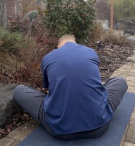
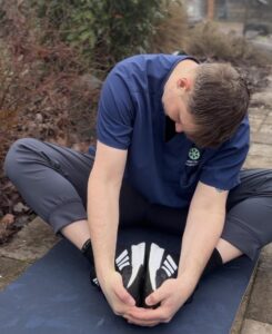
An example of doing a static stretch and incorporating connective tissue techniques would be a traditional standing forward bend. This can stretch your calves and hamstrings. As you incorporate the breath and breathe-in, you will notice your lower abdomen expand and it will stretch your back. Then from your back as you breathe into your chest you can tuck to your chin and stretch your neck creating a line of tension from your toes to the top of your head.
Use the breath to support movements that are complex or threaten the spine. Breathing in to create abdominal pressure to support the spine as you move until you are safe then exhale.
As you breathe deeply and steadily, you may notice an ebb and flow of you stretching that mirrors the tide of your breath. As you inhale, your muscles tighten slightly, reducing the stretch. As you exhale, slowly and completely, your abdomen moves back toward your spine, the muscles in your lower back seem to grow longer, and you can drop your chest toward your thighs.
It’s obvious that exhalation deflates the lungs and lifts your diaphragm into the chest, thereby creating space in the abdominal cavity and making it easier to bend the lumbar spine forward. (Inhalation does the opposite, filling the abdominal cavity like an inflating balloon, making it difficult to fold your spine forward completely.) But you may not realize that exhalation also actually relaxes the muscles of your back and tilts your pelvis forward.
How to Stretch
Warming Up
Stretching is not warming up! It is, however, a very important part of warming up. Warming up is quite literally the process of “warming up” (i.e., raising your core body temperature). Warm up combined with stretching increases the range of motion greater than stretching alonge. A proper warm-up should raise your body temperature by one or two degrees Celsius (1.4 to 2.8 degrees Fahrenheit) and is divided into three phases:
- General warm-up
- Stretching
- Sport-specific activity
General Warm-Up
The general warm-up is divided into two parts: instead of hitting the treadmill or grabbing the weights, try using shaking and patting to safely and gently raise your body temperature.
The best example of a vibration-type exercise is the shaking and patting you see top athletes do before a race or competition. Shaking out, and patting down, a muscle group or part of your body gets it engaged in much the same way as more strenuous exercise. Vibration and patting creates a mechanical force that stimulates endothelial cells to release Nitric Oxide which increases blood circulation.
Start by shaking your hand and feet with small gentle movements. Then incorporate your knees and elbows then move your hips and shoulders and a last twist your spine side to side. Afterwards raise up onto your toes and drop to your heels.
Then you want to pat your skin and connective tissue. Start up your body and down your back. There should be no pain, just your hands and skin should get slightly pink.
These two activities should be performed in the order specified:
- Warming up body
- Joint rotations
Joint Rotations
The general warm-up should begin with joint-rotations, starting either from your toes and working your way up.. This facilitates joint motion by lubricating the entire joint with synovial fluid. Such lubrication permits your joints to function more easily when called upon to participate in your athletic activity. You should perform slow circular movements, both clockwise and counter-clockwise, until the joint seems to move smoothly. You should rotate the following (in the order given, or in the reverse order):
- Fingers, Toes
- Wrists, Ankles
- Elbows, Knees
- Shoulders, Hips
- Neck, Trunk/waist
Warm-Up Stretching
The stretching phase of your warm up should consist of two parts:
- Static stretching
- Dynamic stretching
It is important that static stretches be performed before any dynamic stretches in your warm-up. Dynamic stretching can often result in overstretching, which damages the muscles. Performing static stretches first will help reduce this risk of injury and mitigate any performance decrease.
Static Warm-Up Stretching
Once the general warm-up has been completed, the muscles are warmer and more elastic. Immediately following your general warm-up, you should engage in some slow, relaxed, static stretching.
Dynamic Warm-Up Stretching
Once you have performed your static stretches, you should engage in some light dynamic stretching: leg-raises, and arm-swings in all directions (see section Dynamic Stretching). According to Kurz, you should do “as many sets as it takes to reach your maximum range of motion in any given direction”, but do not work your muscles to the point of fatigue. Remember — this is just a warm-up, the real workout comes later.
Some people are surprised to find that dynamic stretching has a place in the warm-up. But think about it: you are “warming up” for a workout that is (usually) going to involve a lot of dynamic activity. It makes sense that you should perform some dynamic exercises to increase your dynamic flexibility.
Sport-Specific Activity
The last part of your warm-up should be devoted to performing movements that are a “watered-down” version of the movements that you will be performing during your athletic activity. HFLTA says that the last phase of a warm-up should consist of the same movements that will be used during the athletic event but at a reduced intensity. Such sport-specific activity is beneficial because it improves coordination, balance, strength, and response time, and may reduce the risk of injury.
Duration, Counting and Repetition
One thing many people seem to disagree about is how long to hold a passive stretch in its position. Various sources seem to suggest that they should be held for as little as 10 seconds to as long as a full minute (or even several minutes). The truth is that no one really seems to know for sure. According to HFLTA there exists some controversy over how long a stretch should be held. Many researchers recommend 30-60 seconds. For the hamstrings, research suggests that 15 seconds may be sufficient, but it is not yet known whether 15 seconds is sufficient for any other muscle group.
A good common ground seems to be about 20 seconds. Children, and people whose bones are still growing, do not need to hold a passive stretch this long (and, in fact, Kurz strongly discourages it). Holding the stretch for about 7-10 seconds should be sufficient for this younger group of people.
A number of people like to count (either out loud or to themselves) while they stretch. While counting during a stretch is not, by itself, particularly important @dots{} what is important is the setting of a definite goal for each stretching exercise performed. Counting during a stretch helps many people achieve this goal.
Many sources also suggest that passive stretches should be performed in sets of 2-5 repetitions with a 15-30 second rest in between each stretch.
When to Stretch
The best time to stretch is when your muscles are warmed up. If they are not already warm before you wish to stretch, then you need to warm them up yourself, usually by performing some type of brief aerobic activity. Obviously, stretching is an important part of warming-up before, and cooling-down after a workout. If the weather is very cold, or if you are feeling very stiff, then you need to take extra care to warm-up before you stretch in order to reduce the risk of injuring yourself.
Many of us have our own internal body-clock, or circadian rhythm as it is more formally called: Some of us are “early morning people” while others consider themselves to be “late-nighters”. Being aware of your circadian rhythm should help you decide when it is best for you to stretch (or perform any other type of activity). Gummerson says that most people are more flexible in the afternoon than in the morning, peaking from about 2:30pm-4pm. Also, according to HFLTA, evidence seems to suggest that, during any given day, strength and flexibility are at their peak in the late afternoon or early evening. If this is correct then it would seem to indicate that, all else being equal, you may be better off performing your workout right after work rather than before work.
Early-Morning Stretching
On the other hand, if your activity of choice requires considerable flexibility with [little or] no warm-up, you ought to make early morning stretching a part of your routine.” Making your stretching routine as similar as you can to your activity will provide you with the best results In order to do this properly, you need to first perform a general warm-up. You should then begin your early morning stretching by first performing some static stretches, followed by some light dynamic stretches. Basically, your early morning stretching regimen should be almost identical to a complete warm-up. The only difference is that you may wish to omit any sport-specific activity (see section Sport-Specific Activity), although it may be beneficial to perform it if you have time.
Stretching to Increase Flexibility
When stretching for the purpose of increasing overall flexibility, a stretching routine should accomplish, at the very least, two goals:
- To train your stretch receptors to become accustomed to greater muscle length.
- To reduce the resistance of connective tissues to muscle elongation.
If you are attempting to increase active flexibility, you will also want to strengthen the muscles responsible for holding the stretched limbs in their extended positions.
Before composing a particular stretching routine, you must first decide which types of flexibility you wish to increase, and which stretching methods are best for achieving them. The best way to increase dynamic flexibility is by performing dynamic stretches, supplemented with static stretches. The best way to increase active flexibility is by performing active stretches, supplemented with static stretches. The fastest and most effective way currently known to increase passive flexibility is by performing PNF stretches.
Don’t try to increase flexibility too quickly by forcing yourself. Stretch no further than the muscles will go without pain.
Pain and Discomfort
If you are experiencing pain or discomfort before, during, or after stretching or athletic activity, then you need to try to identify the cause. Severe pain (particularly in the joints, ligaments, or tendons) usually indicates a serious injury of some sort, and you may need to discontinue stretching and/or exercising until you have sufficiently recovered.
Common Causes of Muscular Soreness
If you are experiencing soreness, stiffness, or some other form of muscular pain, then it may be due to one or more of the following:
Torn Tissue
Overstretching and engaging in athletic activities without a proper warm-up can cause microscopic tearing of muscle fibers or connective tissues. If the tear is not too severe, the pain will usually not appear until one or two days after the activity that caused the damage. If the pain occurs during or immediately after the activity, then it may indicate a more serious tear (which may require medical attention). If the pain is not too severe, then light, careful static stretching of the injured area is supposedly okay to perform. It is hypothesized that torn fibers heal at a shortened length, thus decreasing flexibility in the injured muscles. Very light stretching of the injured muscles helps reduce loss of flexibility resulting from the injury. Intense stretching of any kind, however, may only make matters worse.
Metabolic Accumulation
Overexertion and/or intense muscular activity will fatigue the muscles and cause them to accumulate lactic acid and other waste products. If this is the cause of your pain, then static stretching, isometric stretching, or a good warm-up or cool-down will help alleviate some of the soreness. Massaging the sore muscles may also help relieve the pain. It has also been claimed that supplements of vitamin C will help alleviate this type of pain, but controlled tests using placebos have been unable to lend credibility to this hypothesis. The ingestion of sodium bicarbonate (baking soda) before athletic activity has been shown to help increase the body’s buffering capacity and reduce the output of lactic acid. However, it can also cause urgent diarrhea.
Muscle Spasms
Exercising above a certain threshold can cause a decreased flow of blood to the active muscles. This can cause pain resulting in a protective reflex which contracts the muscle isotonically. The reflex contraction causes further decreases in blood flow, which causes more reflex contractions, and so on, causing the muscle to spasm by repeatedly contracting. One common example of this is a painful muscle cramp. Immediate static stretching of the cramped muscle can be helpful in relieving this type of pain. However, it can sometimes make things worse by activating the stretch reflex, which may cause further muscle contractions. Massaging the cramped muscle (and trying to relax it) may prove more useful than stretching in relieving this type of pain.
Acknowledgements
Many portions of document rely heavily upon the information in the following books:
- Sport Stretch, by Michael J. Alter(referred to as M. Alter in the rest of this document)
- Stretching Scientifically, by Tom Kurz (referred to as Kurz in the rest of this document)
- SynerStretch For Total Body Flexibility, from Health For Life (referred to as SynerStretch in the rest of this document)
- The Health For Life Training Advisor, also from Health For Life (referred to as HFLTA in the rest of this document)
- Mobility Training for the Martial Arts, by Tony Gummerson (referred to as Gummerson in the rest of this document)
 Willard Sheppy is a writer and healthcare practitioner who seamlessly melds scientific knowledge with practical applications in engaging and authoritative articles. He holds a Bachelor of Science in Environmental Science from Oregon State University and a Master’s in Acupuncture and Oriental Medicine from the distinguished Oregon College of Oriental Medicine.